
IMSE is an inclusive community developing new research paradigms and curricula that bring together engineering, science and medicine to advance personalised precision medicine and train people to seamlessly connect these historically separate fields.
Research focus
projects
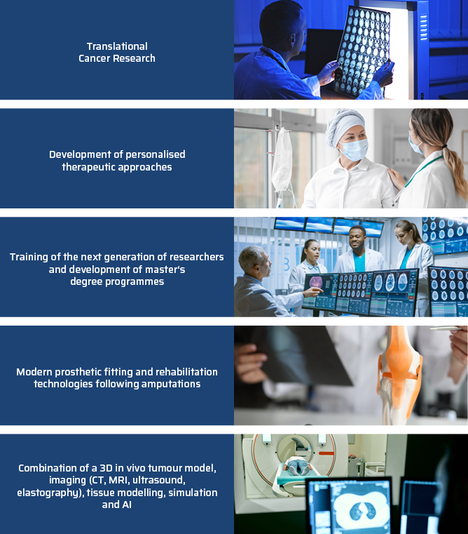
The IMSE’s distinctive profile lies in the close and active collaboration between medical professionals, engineers and other specialist disciplines involved in patient care. It serves as an umbrella organisation and platform for interdisciplinary exchange. Thanks to close links with hospitals and industrial partners, research findings are rapidly translated into practical applications.
Current projects:
ERMES: Information transfer between medical doctors and implanted medical devices via artificial molecular communication
Duration: 01/04/2025 – 31/03/2028
The aim of the project is to develop a novel concept for information transfer for active implantable medical devices. During the meeting, initial research work was presented, focusing on the characterisation of synthetic molecular communication channels and the development of suitable transmitter and receiver systems.
MICROSURGERY MODEL 2: Alternative Methods: Innovative test platform for molecular communication and microsurgical training (Microsurgery Model 2) – Development of novel prostheses
Duration: 1 April 2024 – 30 September 2026
The project aims to utilise the CAM (chorion-allantois membrane) model (1) in microsurgical training for medical students and doctors by incorporating an innovative fluorescence system, (modified) vascular coupling systems and high-frequency ultrasound measurements, and (2) to establish it as an alternative to animal testing, as well as a test platform for innovative technologies and new sensor systems, serving as an innovative prosthetic system in the field of health monitoring.
PANCREATIC CANCER: Interactive patient-specific 3D tumour model for solid tumours and circulating tumour stem cells from pancreatic cancer patients
Duration: 01/07/2024 – 30/06/2027
Objective: AI-based evaluation of histopathological markers in ductal adenocarcinoma of the pancreas before and after treatment with potential chemotherapies in a 3D in vivo tumour model
3D Cysts: 3D in-ovo model for investigating and modulating the growth of human renal cyst tissue and mouse kidney sections – Transregio
Duration: 01/01/2023 – 31/12/2026
The aim of our research is to bridge the gap between animal model results and human studies by testing promising drugs on human ADPKD tissue in ovo.
Partnerships
Innovative research into molecular communication
Prof. Dr Thiha Aung from ISME (THD) has been working closely with Friedrich-Alexander University Erlangen-Nuremberg (FAU) for several years. Together, they are conducting research into the pioneering field of molecular communication.
Natural models and technical approaches
Molecular communication is an innovative, interdisciplinary field of research in telecommunications. Traditionally, electromagnetic waves are used to transmit information, for example in mobile communications.
However, in very small systems at the nano- and micrometre scale, or in liquid media such as blood vessels or oil pipelines, these methods reach their limits.
In nature, however, there are numerous communication systems that function reliably in such complex environments – for example, inter- and intracellular communication or synaptic signal transmission between nerve cells.
Inspired by these natural processes, molecular communication develops synthetic systems in which information is represented by the properties of molecules.
Funded research projects
Prof. Dr Aung is collaborating with Dr Maximilian Schäfer from the Chair of Digital Transmission at FAU on three interdisciplinary projects. These are funded by:
- German Research Foundation (DFG)
- Federal Ministry of Education and Research (BMBF)
- European Union (‘European Innovation Council’)
The projects are developing new concepts for communication within the human body – both theoretically and experimentally – using the CAM model (Chorioallantois Membrane model, a biological test system).
The objectives are:
- Innovative concepts for monitoring patients’ health
- Targeted drug delivery
- Development of new technologies for future medical applications
Through their interdisciplinary research, Prof. Dr Aung and his team are making an important contribution to a better understanding of communication within the body – and thus to the development of groundbreaking medical innovations for the future.
Current Student projects
Abstract:
Hepato-pancreato-biliary (HPB) tumors exhibit high aggressiveness, invasiveness, and therapy resistance. Despite advancements like mFOLFIRINOX, survival rates remain low, and optimal treatment strategies are still undefined. This study integrates AI-based analysis and a 3D in-vivo tumor model to assess the effect of potential therapeutics on histopathological markers. The patient-specific chorioallantoic membrane (CAM) model offers an alternative to animal testing, enabling the cultivation of human tissues and drug testing. For the first time, a combination of Sunitinib and Gemcitabine will be tested in the CAM model, building on prior mouse studies demonstrating superior antitumor efficacy. Histological and immunohistochemical analyses will be performed using markers such as Ki-67, p53, CD3, and CD20 to investigate the proliferation rate, mutation rate, immune response and tumor stroma ratio. AI-assisted tools like QuPath will be employed to enhance digital pathology and automate biomarker quantification. This approach aims to optimize specificity, reduce manual correction efforts, and improve
prognostic assessments. The findings will contribute to a better understanding of therapeutic efficacy, resistance mechanisms, and the potential for personalized treatments.
Abstract:
Die autosomal dominante polyzystische Nierenerkrankung (ADPKD) ist die häufigste Form einer monogenetischen Nierenerkrankung. Die Krankheit ist durch eine fortschreitende, bilaterale Entwicklung und Vergrößerung von Zysten gekennzeichnet, wodurch umliegendes Gewebe verdrängt wird. Infolgedessen benötigen etwa 50 % der ADPKD-Patienten bis zum Alter von 55 Jahren eine Nierenersatztherapie. Da es sich bei ADPKD um eine eher langsam fortschreitende Krankheit handelt, die sich oft über Jahrzehnte entwickelt, bleibt ein großer Zeitrahmen, um das Zystenwachstum zu hemmen und die Nierenfunktion zu erhalten. ADPKD wird hauptsächlich durch heterozygote Mutationen des PKD1- oder
PKD2-Gens verursacht. Die genauen molekularen Mechanismen sind noch nicht vollständig geklärt. Ein besseres Verständnis der Bildung und des Wachstums von Nierenzysten bei ADPKD wäre für die Umsetzung neuer Behandlungsziele und Strategien zur Erhaltung der Nierenfunktion bei diesen Patienten von großem Wert. Derzeit gibt es keine ausreichenden Therapieansätze, um das Zystenwachstum zu inhibieren oder zu verlangsamen und so die Nierenfunktion bei der ADPKD zu erhalten. Der Bedarf solcher Therapieansätze ist aber sehr hoch. Der für die Therapie von ADPKD verwendete Vasopressin 2 Rezeptor Antagonist Tolvaptan (Jinarc®), ist bislang die einzige und auch nur in einigen Ländern
zugelassene Substanz zur Hemmung des Zystenwachstums. Da der Effekt auf das Zystenwachstum nur schwach ausgeprägt ist und von relevanten Nebenwirkungen begleitet wird, ist die Suche nach alternativen therapeutischen Ansätzen unumgänglich. Ein bekanntes Problem bei ADPKD ist die Übertragung von Ergebnissen aus nicht-humanen Modellen auf den Menschen. Das 3D-in-vivo-Modell (Chorioallantoismembran-Modell) kann als Zwischenschritt zwischen Tierversuchen und Studien am Menschen dienen. Als wesentlicher Zwischenschritt zu einer
potentiellen klinischen Anwendung sollen daher vielversprechende Inhibitoren an humanem Zystengewebe in einem 3D-Modell getestet werden. Die Verwendung von menschlichem Zystengewebe stellt nicht nur einen weiteren Schritt in Richtung Translation dar, sondern trägt auch zur Implementierung der 3R-Prinzipien („reduce, refine, replace“) durch Einsatz von Alternativmethoden zum Tierversuch bei.
Abstract:
Pancreatic ductal adenocarcinoma (PDAC) and cholangiocellular carcinoma (CCA) share aggressiveness, short survival time and resistance to chemotherapy. FOXO1 and microRNA-21 function respectively as tumor suppressor and oncogene in PDAC. Moreover, FOXO1 has an important role in vascular homeostasis because it is a fundamental modulator of the formation and maturation of blood vessels.
The cell line with the lowest FOXO1 expression will be selected for transfection. A first transfection experiment will involve the overexpression of FOXO1. Once the transfection is confirmed through western blot and genomic DNA genotyping, the clone will be inoculated onto the CAM. IKOSA will be used to monitor the vessels in the CAM. Part of the explanted cell pellets will be used to analyse the expression angiogenic factors and for immunohistochemistry for the staining of FOXO1 and phospho FOXO1Ser256 (phosphorylated FOXO1). As part of preliminary work, the protein expression of FOXO1 was detected using Western blotting in different PDAC and CCA cell lines. The expression of FOXO1 was significantly higher in BxPC-3 than in MiaPaCa2. Therefore, MiaPaCa2 is a good candidate for overexpression of the gene, while BxPC-3 is a good candidate for knockdown experiments. In addition, cholangiocellular carcinoma tissue was cultured on the CAM model for the first time and the macroscopic course was closely analyzed over a period of one week. The vascularization of CCA onto the CAM was confirmed through ultra-high-frequency ultrasound measurements and we observed the proliferation of the tissue after growth on the CAM using Ki67 staining. The aims are investigating the role of FOXO1 and microRNA-21 in angiogenesis of PDAC and CCA; and cultivating dCCA tumor tissue on the CAM for testing chemotherapeutic drugs.
team


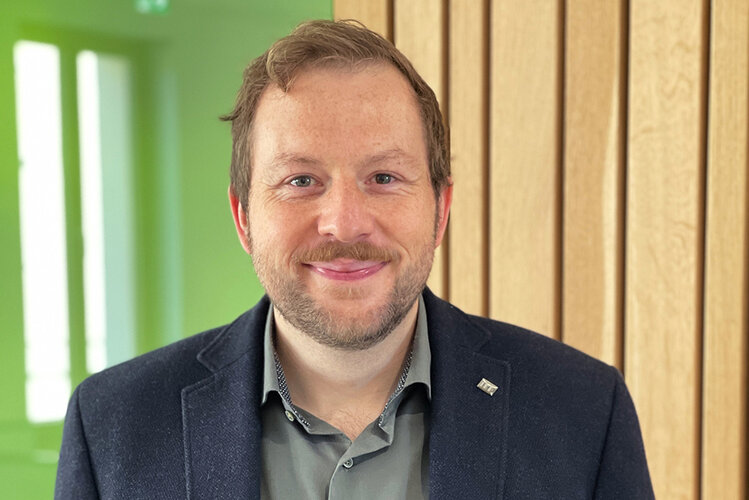

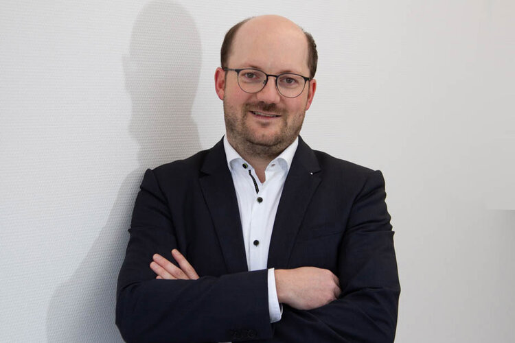
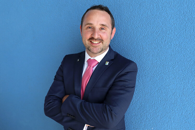
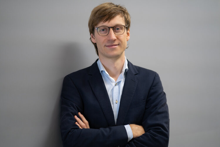





- Agata Montagner
- Laura Lemberger
- Jan Schüler
- Tobias Dobmeier
- Annika Schickle
- Corinna Schütz
- Barbara Silva Salla
- Florian Lang
- Daniel Annang
- Laura Edlbauer
- Emma Meindl
- Janina Poxleitner
- Michelle Fink
- Christiane Loibl
- Tanja Scholz
News
Directions
THD - Technische Hochschule Deggendorf
Campus Hutthurm
Hochleiten 1
94116 Hutthurm
E-Mail: info.tc-hutthurm@th-deg.de